SingaporeSG
 ChinaCN
 MalaysiaMY
 IndonesiaID
 MyanmarMM
Material Science & Nanotechnology
3D Optical Microscopes

UP-5000 3D Optical Profilometer
LARGE SAMPLE IMAGING
MADE EASY
Modular Large Area Optical Microscope UP-5000
6-in-1 Optical Profilometry Imaging Analysis.
Interferometer + Confocal + Spectral Film Thickness + Dark Field + Variable Focus + AFM + Raman
Versatile and Precise Metrology
The optical profilometer UP-5000’s technology, performance, resolution, and versatility creates a class of its own. Equipped with fast scanning, multiple imaging techniques and high resolution, the UP-5000 offers the best solution for large sample surface imaging.
|
High Speed CameraIndustry leading camera with 200 FPS. Quick sub nm precision Measurements. |
Highest Z ResolutionAdvanced latest generation encoders provide the best Z resolution independent of scanning distance or magnification used. |
Modular Versatile PlatformThe 300 x 300 mm high precision cross roller XY stage to fit any sample –wafers, devices, pellicles, components, coupons, etc. |
Powerful Analysis SoftwarePrecise, quantitative, and ISO-compliant analysis software for nanometer resolution studies. |


Why 3D Optical Profilometers?Rtec Instruments optical profilers’ revolutionary surface profiling analyzes any surface with ease. Magnify images and measure profilometry features from nm to mm on a single platform. As a result, our optical systems investigate atomic structures with a single click. Furthermore, our optical profilers measure roughness, waviness, film thickness, and chemical property map. This comprehensive surface profiling approach is the first in the industry. |
|

We Deliver
High Quality
ISO Compliant Metrology Analysis
![]()
Instruments come with standard test protocols to ensure normalized profilometry testing.
Surface Analysis For Every Sample and Situation
|
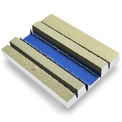
Micro Fluidic Channels |
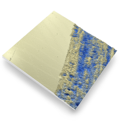
Transparent Coating |
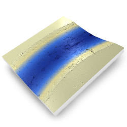
Polymer Coating |
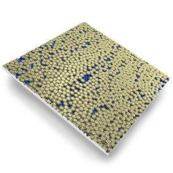
Sub-Nanometer 3D FeaturesRtec 3D Optical Microscopes analyze nm features on rough, transparent, or smooth surfaces. We provide the highest Z and XY optical profilometry resolutions. Additionally, confocal microscopy and white light interferometry are on the same platform. |
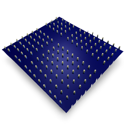
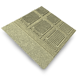
Wafers and SemiconductorsWith stages up to 300×300 mm, analyze full wafer, devices, and pellicles. Check for defects, structures, step height, thickness, particles, and more. Combine AFM, Raman, and 3D optical profilometer data for the same area. |
Features |
Particles |
Wafers |
|
Turning Sample |
Scratch |
Indentation |
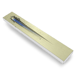
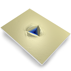
Quality ControlFind cracks and defects with one click. Our 3D optical profilers provide automatic reports and pass-fail criteria analysis. Rtec metrology platforms are the solution for R&D and quality control environments. |

What Are 3D Optical Microscopes?
3D optical microscopes characterize three-dimensional surface topography. First, they optically capture multiple surface images. Then, the microscope stitches the images together in XYZ axis. Finally, they quantifying the data to calculate roughness, step height, film thickness, curvature, bow, and defects. However, all this is not possible with traditional 2D optical microscopy. Our 3D optical profilers use a combination of techniques to provide a quantitative 3D surface. Learn more about these techniques below.
|
What Is Nipkow Confocal Imaging?Confocal microscopy is a non-contact optical imaging technique. It allows for higher resolution and contrast of surface topography utilizing pin holes. First, these pin holes block out-of-focus light during image scanning. Then, white light and a Nipkow confocal disk capture a large sample area at a very high speed creating a three-dimensional scan. The UP-5000 and UP-3000 3D optical microscopes have this feature. |
What Is A White Light Interferometer?White light interferometry (WLI) is a non-contact optical method. WLI characterizes surface topography using scanning interferometry. The technique involves a beam splitter that splits a light beam into a measurement beam and a reference beam. Then these beams are then recombined to create an interference pattern and are then analyzed to generate 2D and 3D models of surfaces. Rtec Instruments’ UP-2000 WLI Optical Profilometers specialize in this feature. |
|
What is Dark Field Imaging?Dark field microscopy is a technique that creates high resolution contrast in specimens that are not imaged well under normal bright field imaging conditions. Using special dark field objectives and a dedicated light system, a dark background is created. This allows for a high degree of contrast for surface features. All Rtec 3D Optical microscopes provide dark field microscopy. |
What Is Variable Focus Imaging?Variable Focus Imaging is an imaging technique used to get a surface image completely in focus. Our microscopes complete this by capturing images at different X, Y, and Z values. Then they stitch the images together to create a fully focused bright field profile of the surface. Both 2D and 3D images use this technique. Each of the Rtec Instruments 3D Optical Microscopes use variable focus imaging for crystal clear imaging. |