 SingaporeSG
 ChinaCN
 MalaysiaMY
 IndonesiaID
 MyanmarMM
Life Science
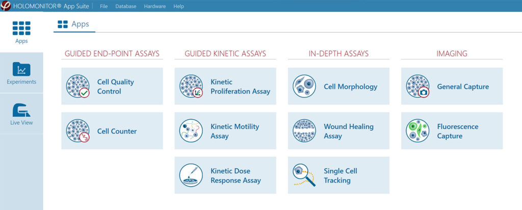
Quantitative Phase Imaging Holographic Monitor

HoloMonitor ® M4
Non-invasive Live Cell Imaging Made Easy
Phase Holographic Imaging provides an innovative tool that lets you continuously image and monitor your cells directly inside your incubator.
HoloMonitor® M4 is a small microscope designed to operate 24/7 inside your normal incubator. Without any labels or stains, it uses digital holography to record your cells in real time. The result is reconstructed 3D images and quantitative data on your cultures, all the way down to single-cell level, collected in a completely non-invasive way.
Cell-friendly, long-term analysis
The HoloMonitor cell culture microscope is an incubator tolerant cell culture imager. This makes it suitable for long-term live cell analysis. Better yet, using quantitative phase-contrast microscopy, it images your cells with no labels or stains. When you place it in a CO2 incubator, you can monitor your cell cultures under optimal conditions. Hence, HoloMonitor allows cell analysis for long periods of time, without having the imaging procedure or instrument affect your cells.
Single-cell analysis on a population level
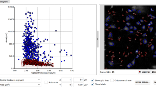
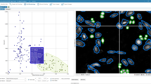
HoloMonitor is a cell culture microscope, which means it images and analyzes cell cultures. However, each image it takes contains data on more than 30 cellular parameters on every single cell. Therefore, you can analyze your cell cultures not only at a population level but down to the single-cell level. The time-lapse images are automatically analyzed as your experiment goes on. Hence, it gives you graphs and scatterplots on single-cell and population data in real-time.
Reuse your cells after the experiment
HoloMonitor does not need labeling, stained or modified cells to be able to image them. The low-energy laser keeps phototoxicity to a minimum, and it images the cells in their normal environment. This means your cells are completely untouched after your HoloMonitor experiment is finished. Therefore, you can use your cells for further studies using other instrumentation, for example, fluorescent imaging by high-resolution confocal microscopy.

Compact to fit inside your standard incubator
The HoloMonitor incubator microscope can be placed on any lab bench. However, it really excels when placed inside a CO2 incubator or hypoxia chamber. It needs only half an incubator shelf, leaving most of your incubator available for regular cell culture work. Further on, HoloMonitor is built to operate 24/7 in a humid environment for long periods of time. This allows you to continuously monitor and analyze your cells, in ideal growth conditions, for as long as you need.
Non-invasive imaging under physiologically relevant conditions
An incubator microscope images your cell cultures where you normally grow them, be it a CO2 incubator or hypoxia chamber. This ensures stable and physiologically relevant growth conditions and minimizes the culture environment’s impact on cell behavior. In addition, HoloMonitor images your cells completely without labels or stains. Hence, you can be sure that the cell behavior you see comes from your experimental treatments—not your lab equipment.
Real-time, single-cell imaging from inside your incubator
HoloMonitor images and analyzes your cells continuously and in real-time, all live from the inside of your standard incubator. Each captured image contains all recorded data for every single cell. This lets you analyze your cell cultures not only on a population level but all the way down to a single-cell level. From the image sequences, you can create time-lapse videos, graphs and scatter plots based on over 30 cellular features. This allows in-depth analysis of your cells’ behavior.
Label-free imaging of living cells means long-term imaging of living cells
The HoloMonitor live cell imaging system uses non-invasive digital holography to monitor your cells. It works completely label-free, uses a low-energy laser, and operates inside your standard incubator. Hence, you can image your cells with a high temporal resolution, for as long as you need, without affecting them. Of course, this is ideal not the least when you work with hard-to-get primary cells. As you do not alter your cells, you can reuse them for other experiments once the imaging is done.
Live cell culture monitoring with HoloMonitor
The HoloMonitor live cell imaging system continuously records multiple time-lapse sequences of your cells right inside your incubator. The image sequences contain a multitude of data, with over 30 cellular features recorded for every single cell. Therefore, it non-invasively gives answers to scientific questions that would normally require you to sacrifice your cells. In essence, HoloMonitor lets you do proliferation, motility, dose-response and wound healing studies without wasting any cells.
Multiple experiments with only one sample
Every image taken with HoloMonitor contains all obtainable data, no matter what assay you were doing while recording it. Hence, you can reanalyze any experiment as a new type of assay. So, if you did a motility assay and need proliferation data on the same cells, just reanalyze your original experiment. This saves time, money and cells. Furthermore, it gives new data on the exact same cells, eliminating variation introduced when preparing new samples with new cells.
Key Benefits
Designed to operate directly inside your incubator
HoloMonitor M4 is designed to operate 24/7, directly inside your normal incubator. You can continuously monitor your cells in their natural environment, making sure you don’t miss any important cellular events in your culture.
Powerful and wide application portfolio
The HoloMonitor App Suite software enables kinetic live-cell tracking and analysis of your cells. In addition to detailed single-cell and cell population data, you get visual data in the form of high-quality images and time-lapse videos. Moreover, you can easily re-analyze the imaging results of previous experiments.
Non-invasive cell imaging with QPI
HoloMonitor is based on digital holography, a QPI technology that quantitatively images your cells with nothing but low-energy light. You don’t need any additives to the media or labels to visualize your cells.
Intuitive, easy-to-use software
HoloMonitor is developed with user-friendliness in mind. Place your cell culture vessel onto the microscope and let the intuitive software guide you from the experimental setup to data analysis.
Gives quantitative data on single-cell level
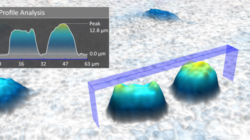
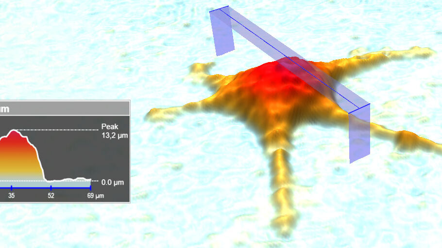
The HoloMonitor App Suite software reconstructs your cells in 3D, immediately providing you with quantitative information about each cell. More than 30 different morphological parameters are calculated automatically, such as cell area, thickness and volume. Of course, all data can be compiled, exported and analyzed at population level as well.
Saves time, money and cells
The HoloMonitor system allows for quick and guided experimental setup and automatically runs the imaging. Furthermore, you can reanalyze experiment data in multiple ways, generating several results from the same cells. This saves you hands-on lab time, money and cells.
Why HoloMonitor?
Cell-friendly
Avoid known interference of cellular markers for biologically relevant results.
Compact
Small footprint to bring imaging inside your incubator.
Affordable
Not all live cell imaging microscopes have to be expensive.
Automated
Get immediate real-time data and image sequences.
Easy-to-use
Intuitive software to guide you from imaging to analysis.
Physiological
Controlled conditions matter for your cell culture.
The HoloMonitor Hardware
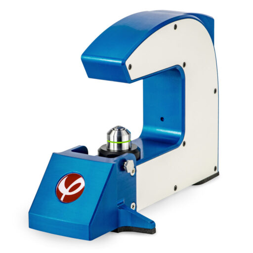
Imaging Unit
The compact base unit is the actual microscope, placed in your incubator together with the motorized stage. It is responsible for continuously imaging your live cell cultures.
| Sample stage | Fixed |
| Light source | External laser unit, 635 nm |
| Sample illumination | 635 nm, 0.2 mW/cm² |
| Lateral resolution | 1 µm |
| Field of view | 0.25 mm² |
| Working distance | 0.5–2.0 mm |
| Autofocusing range | 1.5 mm |
| Maximum image rate | 1 image/s |
| Image size | 1024 × 1024 pixels |
| Dimensions | 290×200×190mm (W × D × H) |
| Weight | 5.15 kg |
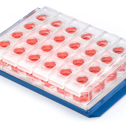
| Cell culture vessels | 6-well, 24-well, 96-well, Petri, IBIDI |
| Power supply | International 110–240 V |
| Interface cable | USB |
| Warranty | 1 year |
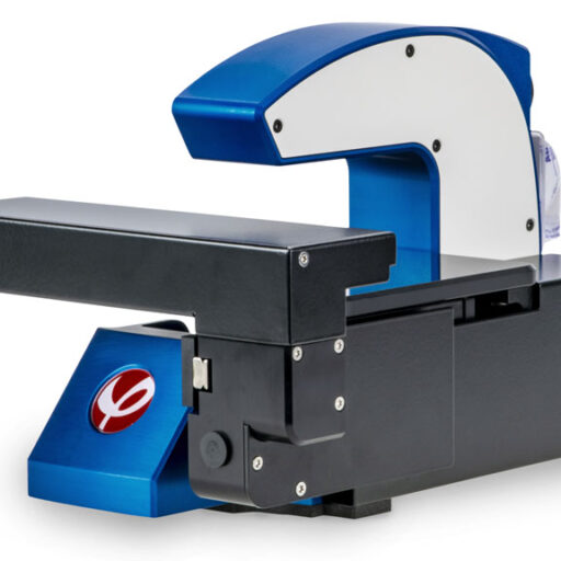
Motorized Stage
The motorized stage supports a variety of culture vessels. Moreover, it enables automated and continuous imaging of multiple positions in your sample.
| Repeatability | 5 µm |
| Travel range | 100 × 70 × 10 mm (X × Y × Z) |
| Dimensions* | 290 × 200 × 185 mm (W × D × H) |
| Operational dimensions* | 375 × 260 × 185 (W × D × H) |
| Minimum space inside the incubator | 400 × 270 × 185 (W × D × H) |
| Weight* | 5.5 kg |
| Operating temperature | 10–40°C |
| Operating humidity | Max 95% |
| Cell culture vessel support | 6-well, 24-well, 96-well, Petri, IBIDI |
| Warranty | 1 year |
*Including the base unit
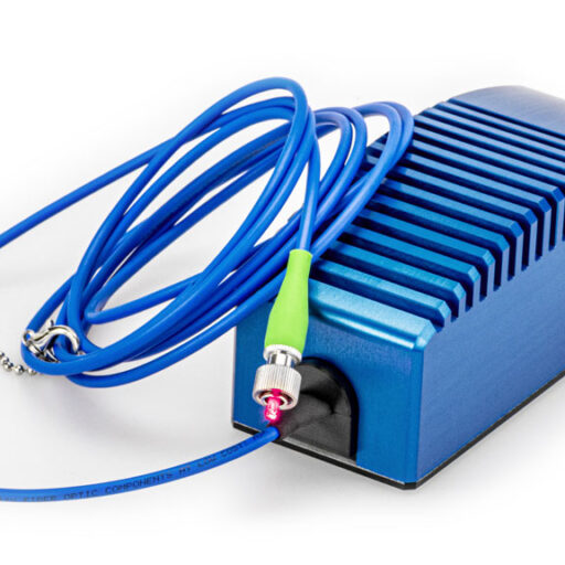
Laser Unit
The laser unit, placed outside your incubator, supplies the system’s low-energy laser for label-free holographic live cell imaging that leaves your cells unaffected.
| Exposure time | 5 ms, non-scanning |
| Light source | External laser unit, 635 nm |
| Sample illumination | 0.2 mW/cm² |
| Power supply | International 110–240 V |
| Interface cable | USB |
| Warranty | 1 year |
The add-on fluorescence unit makes it possible to add a green fluorescence dimension to the label-free holography data and study your cells’ behavior in more detail.
| 1-channel fluorescence | Optimized for EGFP (FITC-Cy2) |
| Excitation | 470 nm filtered LED, with bandwidth (FWHM): 40 nm |
| Emission | 525 nm filtered, with bandwidth (FWHM): 50 nm |
| Exposure time | 1 – 1000 ms |
| Gain | 1 – 4 |
| Magnification | 10x, matched with mounted HoloMonitor M4 |
| Sensor | 1280 x 1024 px, 1.31 MPix |
| Image Resolution | 0.5 µm |
| Field of view | 0.25 mm² |
| Image size | 1024 × 1024 pixels |
| Dimensions | 310 x 180 x 85 mm (W × D × H) |
| Weight | 4.0 kg |
| Power | < 5 W |
| Interface | USB connector |
| Software requirement | HoloMonitor App Suite software 4.0 |
| Operating environment | 10 – 50 °C, < 95 % relative humidity |
Click HERE to learn more.
The HoloMonitor Software
The heart of HoloMonitor is the modular App Suite software, with tailor-made apps for different live cell assays. Each app offers a guided setup of your experiment, automatic image collection and real-time data analysis and presentation.
Moreover, App Suite excels when it comes to data re-analysis. Every experiment contains all the data necessary to reanalyze it as another type of experiment. For example, a proliferation assay can be reanalyzed as a motility assay at a later point. Hence, no need to spend time, money and cells on a new experiment!
The Workflow
Designed with cell biologists, the HoloMonitor App Suite software offers a simple and intuitive user interface, guiding you through the workflow from experiment setup to data presentation in 5 steps.
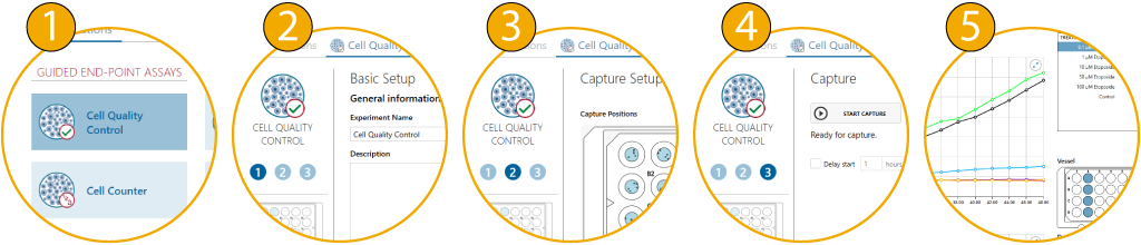
In addition, you can easily create colorful videos and images to present your results. Of course, all data can also be exported to Microsoft Excel for further analysis.
HoloMonitor Accessories
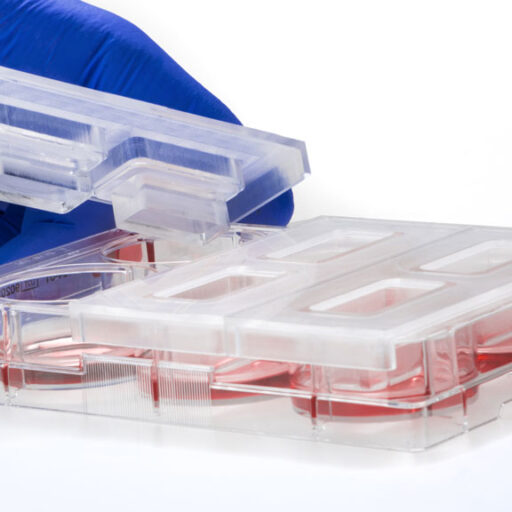
Culture Vessel Holders
HoloMonitor’s motorized stage supports a variety of culture vessels, each secured with a vessel holder. This means easy attachment and exact positioning on the microscope.
HoloLids™
HoloLids are specially designed lids for standard cell culture vessels. They are optically superior to normal vessel lids and ensure the best possible image quality every time.
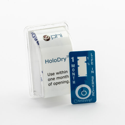
HoloDry™
HoloDry is a non-toxic canister that is easily attached to the HoloMonitor microscope. It keeps the optics and interior free of condensation, which ensures optimal performance.
| Light source | External laser unit |
| Sample illumination | 635nm, 0.2mW/cm2 |
| Objective | 20x |
| Lateral resolution | 1µm |
| Field of view | 567µm × 567µm |
| Working distance | 0.5 – 2mm |
| Autofocusing range | 1.5mm |
| Image capture rate | 1 image/s |
| Image size | 1024 × 1024 pixels |
| Stage travel range | 100×70×10 (X×Y×Z) |
| Stage repeatability | 5µm |
| HoloMonitor® M4 dimensions | 290×200×190mm (W×D×H) |
| Space required in incubator | 400×270×190mm (W×D×H) |
| Weight | 5.15 kg |


Authors: Frida Berlin
Journal: Doctoral thesis (2023)
Research Areas: Cell biology
Keywords: HoloMonitor M4, Mast cells, proteases, tryptase, chymase, chronic respiratory diseases, asthma
Chronic respiratory diseases, such as asthma, are an increaseing health issue worldwide and cause about 3.9 million deaths annually. Despite this, little is know about the molecular mechanisms underpinning disease pathogenesis. Bronchial and alveolar remodeling and impaired epithelial function are typical characteristics of chronic respiratory diseases. In these patients, an increased number of mast cells, positive for the serine proteases; tryptase and chymase, infiltrate the epithelium and the alveolar parenchyma. While it is likely that the epithelial cells are exposed to various amounts of released tryptase and chymase, the interaction between mast cells and epithelial cells remains unknown. This thesis aimed to investigate the impact of mast cell proteases on bronchial and alveolar remodelling. Human bronchial and alveolar epithelial cells were treated with tryptase and chymase. Holographic live cell imaging, fluorescent microscopy, and gene and protein assays were used to analyze various parameters such as proliferation patterns, protein expressions and distributions. The results showed that both tryptase and chymase promoted epithelial remodelling in several ways. Tryptase induced cell growth, cell survival, and wound healing, whereas chymase reduced cell growth, altered cell morphology and impaired epithelial barrier proprties. In conclusion, our results suggest that intraepithelial and alveolar mast cell release of proteases plays a crucial role in epithelial homeostasis, and that an inbalance of the protease release may be involved in respiratory disease progression and in disruption of critical epithelial functions.
Authors: Li et al.
Journal: Experimental Neurology (2023)
Research Areas: Cancer research
Cell Lines: U251, U373, HEK293T, HA
Keywords: HoloMonitor M4, cell motility, Glioblastoma STK24P1, P1-121aa, ELF2 Phosphorylation
Glioblastoma (GBM) is the most common malignant tumor of the central nervous system. Vasculogenic mimicry (VM) is a hematological system composed of tumor cells that exert blood perfusion without relying on vascular endothelial cells. The current poor results of anti-vascular therapy for clinical GBM are associated with the presence of VM; therefore, it is important to investigate VM formation in GBM. The results demonstrate thatSTK24P1 encodes P1-121aa with a kinase structural domain, and in vitro kinase assays demonstrated that P1-121aa mediates modification of ELF2 phosphorylation. ChIP and dual luciferase reporter gene assays demonstrated that the transcription factor ELF2 binds to VE-cadherin and the VEGFR2 promoter region, thereby promoting VM formation in glioma cells. P1-121aa, encoded by the pseudogene STK24P1, phosphorylates ELF2 at S107, increasing the stability of the ELF2 protein. ELF2 promotes VEGFR2 and VE-cadherin expression at the transcriptional level, which in turn promotes VM in GBM. This study demonstrates the important roles of STK24P1, P1-121aa, and ELF2 in regulating VM in GBM, which could provide potential targets for GBM treatment.HoloMonitor M4 is used to study the effect of different mutations on the migration ability of human glioma cell.
Authors: Wasilewska et al.
Journal: International Journal of Biological Macromolecules (2023)
Research Areas: Materials Science
Cell Lines: MC3T3-E1
Keywords: HoloMonitor M4, Cell proliferation, Cell morphology, Macroion multilayers, Polysaccharide layers, Label-free biosensors, Streaming potential, OWLS, Resonant waveguide grating, Cell adhesion, Antimicrobial coatings, QCM
The regulation of cellular adhesion is a crucial aspect in the development of biomaterials and cell-based biosensing assays. In this study, synthetic poly(diallyldimethylammonium chloride) (PDADMAC), natural chitosan, and heparin were utilized to assemble PDADMAC/heparin and chitosan/heparin films. The physicochemical properties of these macroion multilayers were characterized using streaming potential measurements (SPM), quartz crystal microbalance (QCM-D), and optical waveguide lightmode spectroscopy (OWLS), while their topography was imaged using atomic force microscopy (AFM). The adhesion of the preosteoblastic cell line MC3T3-E1 on these well-characterized polysaccharide-based multilayers was evaluated using a resonant waveguide grating (RWG) based optical biosensor and digital holographic microscopy HoloMonitor M4. Results showed that PDADMAC/heparin films were the most effective in inducing cellular adhesion, while chitosan/heparin-based multilayers exhibited negligible cell attachment. These findings suggest that polysaccharide-based multilayers have potential for use in medical applications.HoloMonitor M4 is used to study the cell proliferation and morphological parameters of preosteoblastic cells on different coatings.
Authors: Frida Berlin et al.
Journal: Cells (2023)
Research Areas: Cell Biology
Cell Lines: BECs, BEAS-2B, AECs, A549
Keywords: HoloMonitor M4, cell proliferation, mast cell, proteases, tryptase, bronchial epithelium, alveolar epithelium, growth factors, anti-apoptosis, airway remodeling, cell growth, par-2
The article investigates the role of mast cell tryptase in bronchial and alveolar remodeling and the mechanisms of regulation during inflammation. The study found that mast cell tryptase enhanced human bronchial and alveolar epithelial cell growth and shortened the cell division intervals. The elevated cell growth induced by tryptase remained in a pro-inflammatory state. Tryptase also increased the expression of the anti-apoptotic protein BIRC3, as well as growth factor release in epithelial cells1. HoloMonitor is used to investigate the cell growth and cell division effects of mast cell tryptase on bronchial and alveolar epithelial cells.