CATALOG NUMBER | UNIT SIZE |
FN10100 |
FluoMag-N 100 µL |
FC10100 |
FluoMag-C 100µL |
FP10100 |
FluoMag-P 100µL |
FS10100 |
FluoMag -S 100µL |
FV10100 |
FluoMag-V 100µL |
RECOMMENDED FOR:
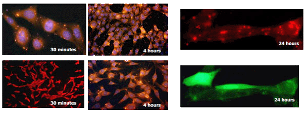
Figure 1: NIH-3T3 transfection with FluoMag-P and pEGFP plasmid DNA. NIH-3T3 cells (5x10⁴ cells/well), growing on coverslips, were transfected in 24-well plates with 1 µg of pEGFP plasmid and 1µL of FluoMag-P per well as described in the Magnetofection instruction manual. At several times post-transfection, cells were fixed, stained with DAPI to detect nucleus (blue) and observed under a fluorescent microscope equipped with a CCD camera.
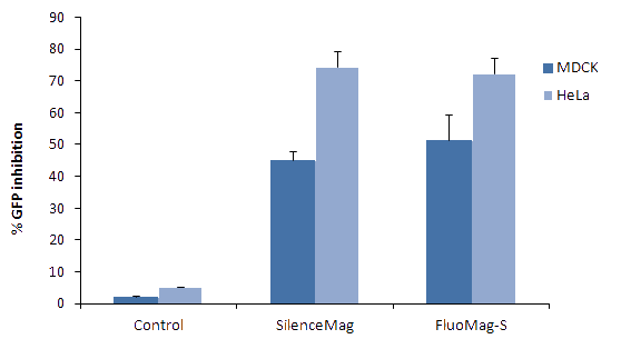
Figure 2: Transfection efficiency of FluoMag-S versus SilenceMag. GFP stably transfected MDCK and HeLa cells were plated the day before transfection in a 24-well plate. Cells were then treated with SilenceMag or FluoMag-S and siRNA (targeting GFP or targeting LacZ as control) as described in the SilenceMag instruction manual. Complexes were prepared with 1 μL of SilenceMag or FluoMag-S and 10nM (67.5ng) of siRNA. Cells were then transfected in 500 μL transfection volume. GFP expression level was monitored 72h post-transfection by detection of fluorescence intensity with a fluorometer.
Technical Resources | Kindly refer to the "Downloads" tab. |
Sauer AM., J Control Release. 2009 Jul 20;137(2):136-45.
Magnetofection, gene delivery under the influence of a magnetic field, is a technique to increase transfection efficiency by enforcing gene vector contact with a target cell. Mechanisms of magnetic lipoplex internalization and intracellular details of magnetofection are still unknown. In this study, cellular dynamics of magnetic lipoplexes were examined in real time by means of highly sensitive dual-color fluorescence microscopy. Single particle tracking of magnetic lipoplexes provided trajectories representing the movement of the lipoplexes during internalization and subsequent intracellular processes. Magnetic lipoplexes show a three-phase behavior similar to polyplexes. During phase I lipoplexes are attached to the cell surface and show slow cooperative transport behavior. Phase II takes place inside the cell and was characterized by anomalous and confined diffusion. Phase III represented active transport along microtubules inside the cell. The majority of lipoplexes were internalized via endocytosis during phase I. On later time scales the formation of a perinuclear ring was observed. Persisting colocalization of fluid phase marker and lipoplexes after 24 h indicated slow endosomal release. In short, the internalization characteristics of magnetic lipoplexes are very similar to that of polyplexes. Furthermore our results suggest that the magnetic field induces an increased concentration of magnetic complexes on the cell surface resulting in higher transfection efficiency.
Al-Shakli AF., 2018 Keele University
Neurons are the targets of injury and disease in many neurological conditions, and achieving neuronal survival/repair is a key goal for regenerative medicine. In this context, genetic engineering of neurons offers a platform for (i) basic research to enhance our understanding of neuronal biology in normal, disease/and injury conditions; and (ii) for regenerative medicine to enhance the functionality of neurons. Although, a wide range of attempts have been made to promote gene delivery to primary neurons, these cells are still difficult to genetically engineer, and current methods rely heavily on viral vectors which pose safety considerations. Magnetic nanoparticles (IONPs) are currently of great interest in regenerative medicine including for non-viral gene delivery by the 'magnetofection' strategy, i.e when used with applied magnetic fields. This project aimed to examine (i) the influence of two novel uniaxial and biaxial oscillating magnetic field devices on primary neuronal transfection efficiency, and (ii) examine the safety of magnetofection using histological and electrophysiological studies. In order to do this, a robust protocol to derive primary cortical neurons was first established.
A second issue is that surgical delivery of Neurons results in low survival. Additionally, most basic research has relied on neurons grown on 'hard‘ substrates such as plastic, which do not mimic the mechanical properties of the in vivo microenvironment. To address these limitations, primary cortical neurons were grown in a 3-dimensional 'soft' collagen hydrogel construct which can serve both as a protective cell delivery system and a 'neuromimetic' substrate. The safety of the established protocol was evaluated by electrophysiological analyses on neurons.
The findings demonstrate that the safety of magnetofection is magnetic field dependent, and at optimal conditions, electrophysiological properties of the nano-engineered neurons were normal. Secondly, I have shown that collagen hydrogels can support the 3D growth of neurons and electrophysiological studies can be carried out on the construct neurons; small differences were found between neurons grown on hard and soft materials. Finally, the amenability of genetic engineering of neurons within hydrogels using IONPs has been shown.
Sign In to Start Your Session